
Retinal Tear and Detachment
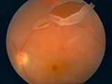 The vitreous is a clear, viscous liquid that fills the central cavity of our eyes and gives them shape. When we are young, the vitreous has a thick, gelatinous consistency and is firmly attached to the retina. As we age, the vitreous liquefies and eventually separates from the retina. Although this usually results in nothing more than a few harmless floaters, tension from the detached vitreous can sometimes tear the retina.
If liquefied vitreous passes through the tear and collects beneath the retina, the retinal tear can become a retinal detachment. Retinal detachment can cause significant, permanent vision loss and requires immediate medical treatment.
There are three kinds of retinal detachments. The most common form (rhegmatogenous retinal detachment), described above, occurs when fluid accumulates beneath the retina. People who are nearsighted or who have had an injury or eye surgery are most susceptible to this form. Less frequently, a pulling force can develop between the retina and vitreous or scar tissue, which may pull the retina loose (tractional retinal detachment). This type of detachment occurs most often in patients with diabetes. Third, disease-related fluid can accumulate under the retina and push it away from the eye wall (serous retinal detachment).
Signs of retinal tear or detachment include flashes of light, a group or web of floaters, wavy or watery vision, a sense that there is a veil or curtain obstructing vision, or a sudden drop in vision quality. If you experience any of these symptoms, call your doctor immediately. Early treatment is essential to preserve your vision.
For more information, we welcome you to view the American Academy of Ophthalmology page on retinal detachment. Please click here.